The Optic Canal A Critical Yet Often Overlooked Brain Highway
The Optic Canal A Critical Yet Often Overlooked Brain Highway
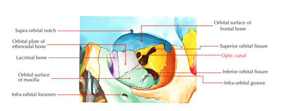
The optic canal, a slender bony passageway in the skull, serves as the indispensable bridge between the eye and the brain—yet remains profoundly underappreciated in both clinical and public understanding. Stretching only about 1.5 centimeters, this narrow channel transmits the optic nerve (cranial nerve II) and the middle meningeal artery, forming a lifeline essential for vision and neurological integrity. Despite its small size, damage or compression at this site can disrupt sight and cognition, underscoring the optic canal’s role as a critical, yet frequently overlooked, conduit in neuroanatomy.
Behind the orbits, nestled between the sphenoid bone and the ethmoid, the optic canal’s precise architecture is both fragile and vital. It allows the optic nerve fibers—responsible for relaying visual information from the retina—to connect seamlessly to the thalamus, specifically the lateral geniculate nucleus, the brain’s primary visual processing center. Disruption at this junction impairs visual signaling, potentially leading to blurry or lost vision, double images, or even cortical vision loss.
At the same time, the middle meningeal artery’s passage through the canal supplies important vascular nourishment; compromise here may compromise tissue health and healing following injury or inflammation.
The Anatomy of Critical Function
The optic canal’s narrow dimensions—approximately 8 mm in diameter—make it a high-risk site for mechanical and vascular stress. Compression or swelling from tumors, fractures, or inflammatory conditions like diabetes-related microvascular changes can constrict the canal and threaten optic nerve function. “The optic canal is not merely a structural access route—it’s a dynamic interface,” notes neuroanatomist Dr.Elena Marco, an expert in cranial nerve pathways. “Its integrity ensures unimpeded transmission of sensory data from the eye to the brain. Damage here doesn’t just cause immediate vision loss; it often signals broader neurological compromise.”
The canal’s role extends beyond sight: its close association with cranial nerve II makes it a linchpin in neurological diagnostics.
Clinicians rely on recognizing signs such as proptosis, optic disc swelling, or visual field deficits—early warnings of intracranial pressure or structural damage—that often trace back to pathology at the canal’s entrance. Yet, when symptoms manifest, the canal’s subtlety often leads to misdiagnosis or delayed treatment, especially in patients without accessible imaging of this microscopic pathway.
Clinical Implications: From Tumors to Trauma
Several conditions underscore the optic canal’s clinical significance. Pituitary tumors, particularly adenomas, frequently impinge on the canal as they expand, compressing the optic nerve and artery.This can produce characteristic visual symptoms including bitemporal hemianopia—a loss of peripheral vision in both eyes—before permanent damage occurs. Similarly, basilar skull fractures, though rare, may fracture the canal’s bone, risking hemorrhage or scarring that jeopardizes neural continuity.
Even benign fluctuations—such as those seen in idiopathic intracranial hypertension—can subtly alter pressure dynamics within the canal, affecting optic nerve perfusion over time.
In younger patients, congenital stenosis—a rare developmental narrowing—poses diagnostic challenges, often mistaken for migraines or retinal issues. Each scenario demands acute awareness from neurologists, ophthalmologists, and neurosurgeons alike.
Diagnostic Tools: Unlocking the Hidden Pathway
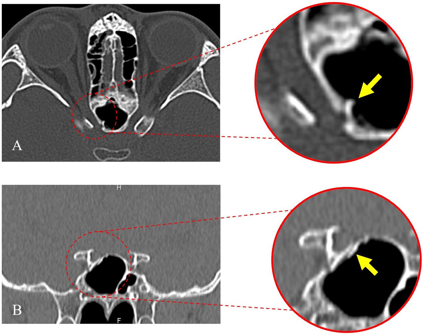
Advances in neuroimaging have dramatically improved identification of lesions within the optic canal. High-resolution MRI with dural window sequences provides clear visualization of both neural and vascular structures, enabling early detection of tumors, vascular inflammation, or microfractures.Angiography further clarifies blood flow patterns, crucial for assessing middle meningeal artery involvement. These tools not only enhance diagnosis but also guide minimally invasive interventions—such as endoscopic decompression or targeted radiofrequency ablation—preserving function where traditional surgery risks collateral damage.
Yet despite such progress, many cases remain underrecognized.
Patients often present with vague symptoms— gradual vision decline, subtle visual field cuts—that tell no immediate story. Radiologists and clinicians must maintain vigilance, especially when optic nerve pathway integrity is at stake. A single missed canal lesion can derail recovery, so multidisciplinary collaboration—between neuroscientists, radiologists, and surgical teams—proves essential for optimal outcomes.
Preserving a Mission-Critical Connection
The optic canal, though microscopic in scale, lies at the heart of one of the brain’s most vital missions: interpreting light and transforming it into sight.Its role as a conduit between eyes and brain is deceptively simple, yet its failure has cascading consequences. Whether due to tumors, trauma, or pressure, disruption here threatens not just vision but cognitive and perceptual stability. Advances in imaging and clinical awareness have begun to correct longstanding oversights, revealing the canal’s true status—as a critical, yet profoundly important brain highway.
Protecting and understanding this pathway is not merely academic—it is essential for safeguarding one of humanity’s most precious senses.


Related Post

WA Time Sets the Rhythm: Current Moment Drives WA’s Pulse in Business, Travel, and Daily Life

ClickHouse Alternatives: Top Competitors That Replicate Its Power for Big Data Needs

Behind the Lens: A Deep Dive into Bella Ramsey’s Parenting in the Spotlight

A Uplifting Journey of Kerri Browitt Caviezel: From Shadows to Starlight


